Identify My Invertebrate?

ziggypie

Axolotl Enthusiast
Joined
Jun 6, 2007
Messages
29
Reaction score
1
Points
0
Location
New York, USA
Country
United States
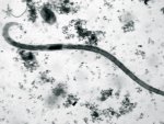
It's odd to say, but this looks like either some sort of Cestode, or a larval form of something much bigger than anything I have in the tank (save the Axolotls and the fake decor). I took this picture from a dissecting scope, I was silly and forgot to record the power, but I think it was between 10 and 20x (obviously no ocular power). If it helps, that blurb that you see next to it is an ostracod crustacean ~0.3 mm. Thank you!
 

Attachments

  • MVI_42411.jpg
    MVI_42411.jpg
    110.5 KB · Views: 264
It's a worm, that's about all I can tell you. Perhaps a nematode? I'm afraid I'm not very good at the game of "name that invert". :cry:

May I use your photo for Caudata Culture? I'm assembling a page of photos of "tank critters", and this would fit right in. Please PM me and tell me what name the copyright credit should be to.
 
I'm fairly certain its not a Cestode. I assume thats the head on the photo which means it appears to lack a means of securing itself inside a host which is characteristic of most Cestodes. At a guess I'd say it was a small Annelid. Do you have a sand based substrate on the bottom? Is it possible for you to post a bigger picture of the whole organism as I could run it past my parasitology lecturers to see if they recognise it.
 
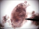
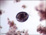
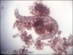
I was having fun taking more pictures, and so let's turn this into a game! I know most of my little friends (except for the worm you will see- as we obviously haven't gotten that one figured out yet, so keep at that one in the first post if you can, haha), but here we go!! ::booming game show voice:: "IDENTIFY MY INVERTEBRATES!" hehehe... And Jennewt, if you like these, too, then they are yours for the benefit of the forum! :)

I forgot to mention that these were all taken with a compound light microscope- under either scanning or low power!
 

Attachments

  • IMG_4272.jpg
    IMG_4272.jpg
    62.1 KB · Views: 213
  • IMG_4285.jpg
    IMG_4285.jpg
    35.7 KB · Views: 215
  • IMG_4288.jpg
    IMG_4288.jpg
    35.2 KB · Views: 203
  • IMG_4299.jpg
    IMG_4299.jpg
    48 KB · Views: 209
  • IMG_4304.jpg
    IMG_4304.jpg
    38.7 KB · Views: 202
  • IMG_4306.jpg
    IMG_4306.jpg
    39.5 KB · Views: 214
  • IMG_4310.jpg
    IMG_4310.jpg
    46 KB · Views: 209
The 5th and 6th are ostracods, those are the only ones I know for sure. Thanks for posting!
 
Correct so far! I love the first picture because if you look inside the sac there is a Rotifer just hanging out, looking so very cool and composed. If you look on another one of my threads, you'll see I have a special place in my heart for my wheel-bearers :) Keep at it, folks!
 
I finally got these photos posted on CC:
http://www.caudata.org/cc/articles/Tank_critters2.shtml

Does anyone care to venture an ID for the first 3 photos in the group above?


P.S. I have moved this thread out of the axolotl area, as it seems to be of more general interest and not axie-specific.
 
Last edited:
Just come across this thread. Nice photos!

The 1st photo looks to me like a ciliate protozoan.

The 2nd is a bdelloid (leech-like) rotifer.

The 3rd looks like another ciliate, though it's hard to be sure.

The 4th is another rotifer.

5 and 6 are ostracods. I vaguely recognise the creature with the ostracod but don't have my book handy I'm afraid.

7 looks like a small oligochaete worm (there are a lot of species and they're tricky to identify - theres a good link at http://sunsite.ualberta.ca/Projects/Aquatic_Invertebrates/?Page=17 )

Tristan
 
With the worm in the first post, I think it's an annelid worm, since it appears to be segmented. Nematodes do not have segmentation. It's possibly an Oligochaete, but I'm not into non-arthropod invertebrate identification.

As a fun trivia, nematodes are eutelic, which means that when they're adults, they have a fixed number of cells in their body.
 
General chit-chat
Help Users
  • No one is chatting at the moment.
  • Katia Del Rio-Tsonis:
    Dear All, I would appreciate some help identifying P. waltl disease and treatment. We received newts from Europe early November and a few maybe 3/70 had what it looked like lesions under the legs- at that time we thought maybe it was the stress of travel- now we think they probably had "red leg syndrome" (see picture). However a few weeks later other newts started to develop skin lesions (picture enclosed). The sender recommended to use sulfamerazine and we have treated them 2x and we are not sure they are all recovering. Does anyone have any experience with P. waltl diseases and could give some input on this? Any input would be greatly appreciated! Thank you.
    +1
    Unlike
  • Katia Del Rio-Tsonis:
    sorry I am having a hard time trying to upload the pictures- I have them saved on my hard drive... any suggestions-the prompts here are not allowing for downloads that way as far as I can tell. Thanks
    +1
    Unlike
    Katia Del Rio-Tsonis: sorry I am having a hard time trying to upload the pictures- I have them saved on my hard... +1
    Back
    Top